When a pet is unwell, knowing exactly what is happening inside the body is essential for effective treatment. Advanced veterinary imaging has transformed how veterinarians diagnose and treat complex conditions, offering a non-invasive window into soft tissues, bones, the brain, and the spinal cord. These technologies play a critical role in modern veterinary medicine, particularly in neurology, oncology, and orthopedics.
What Is Advanced Veterinary Imaging?
Advanced veterinary medical imaging refers to diagnostic technologies that go beyond standard X-rays. While radiographs are useful for evaluating bones and some soft tissue structures, they provide limited detail for the brain, spinal cord, internal organs, and vascular systems. Advanced imaging fills this gap with far greater precision and detail.
The two most commonly used advanced veterinary imaging modalities are Magnetic Resonance Imaging (MRI) and Computed Tomography (CT). Both are widely used in human medicine and have been adapted for use in animals with excellent results.
Types of Veterinary Imaging Services
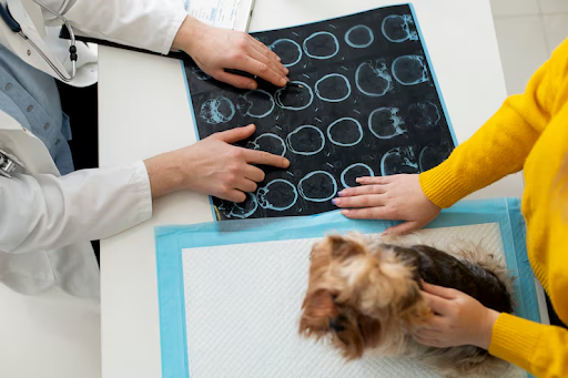
MRI (Magnetic Resonance Imaging)
MRI is the gold standard for imaging the brain and spinal cord in veterinary patients. It uses strong magnetic fields and radio waves — not radiation — to produce highly detailed images of soft tissue structures. MRI is particularly valuable for diagnosing conditions such as intervertebral disc disease, brain tumors, inflammatory brain disease, and spinal cord abnormalities. It is highly sensitive and can detect early-stage lesions that would be invisible on X-ray or CT.
CT (Computed Tomography)
CT scanning uses a series of X-ray images taken from different angles to produce cross-sectional views of the body. It is faster than MRI and is excellent for visualizing bony structures, the lungs, abdominal organs, and complex fractures. CT is often used in trauma cases, to assess the skull and inner ear, and for surgical planning. Some facilities also offer CT angiography to evaluate blood vessels.
Ultrasound
Ultrasound uses high-frequency sound waves to produce real-time images of soft tissue organs such as the heart, liver, kidneys, and bladder. It is particularly valuable for cardiac evaluations (echocardiography), guided biopsies, and assessing abdominal masses. It requires no anesthesia in most cases and provides instant results.
Why Is Advanced Veterinary Medical Imaging Important?
Standard physical examinations and basic bloodwork can identify many health problems, but they have limitations when it comes to the nervous system, internal organs, and complex structural conditions. Advanced imaging allows veterinarians to visualize exactly where a problem is located, how extensive it is, and what structures are involved. This leads to more accurate diagnoses, better treatment planning, and improved patient outcomes.
For veterinary neurology patients in particular, MRI is not a luxury — it is a necessity. Conditions such as IVDD, brain tumors, and inflammatory diseases of the nervous system cannot be reliably diagnosed without it.
What to Expect During a Veterinary Imaging Appointment
Most advanced imaging procedures require the patient to remain completely still, so general anesthesia is typically used. A board-certified veterinary radiologist or specialist will analyze the images and provide a detailed report. The results help guide the treatment plan, whether that involves surgery, medication, radiation therapy, or monitoring.
The anesthesia protocol is carefully tailored to each patient’s age, weight, and overall health to minimize risk.
Is Advanced Imaging Safe for Pets?
Yes. MRI does not use radiation and is considered very safe. CT scanning involves a small amount of radiation, but doses are minimal and risks are low, especially when weighed against the diagnostic benefit. Ultrasound is entirely radiation-free. The primary risk associated with advanced imaging is the anesthesia required to keep the patient still — a risk that experienced veterinary teams are well-equipped to manage.
Konklusion
Advanced veterinary imaging has become an indispensable part of modern pet healthcare. From pinpointing the exact location of a spinal disc herniation to identifying a brain lesion invisible to the naked eye, these veterinary imaging services give specialists the information they need to act decisively. If your veterinarian recommends advanced imaging for your pet, it is a powerful step toward a precise diagnosis and an effective treatment plan.
.For more info visit :- flowactivo.org